Balaton Summer School 2025 offers an immersive experience with acknowledged experts in the field of medical and biological physics. Learn about the latest research and technologies in the field, and connect with fellow physics students from around the world. Don’t miss this opportunity to gain valuable insight into this quickly evolving field that has so much potential to improve health and lives!
BSS will take place in the beautiful city of Balatonfüred, located on the northern shore of Lake Balaton in Hungary. With its stunning views of the lake and surrounding hills, Balatonfüred is a popular tourist destination known for its natural beauty and vibrant culture. Visitors can explore the charming streets of the historic town center, take a dip in the refreshing waters of Lake Balaton, or enjoy local cuisine and wine at one of the many restaurants and wineries in the area. The city is easily accessible by public transportation and offers a comfortable and convenient base for attending the Summer School.
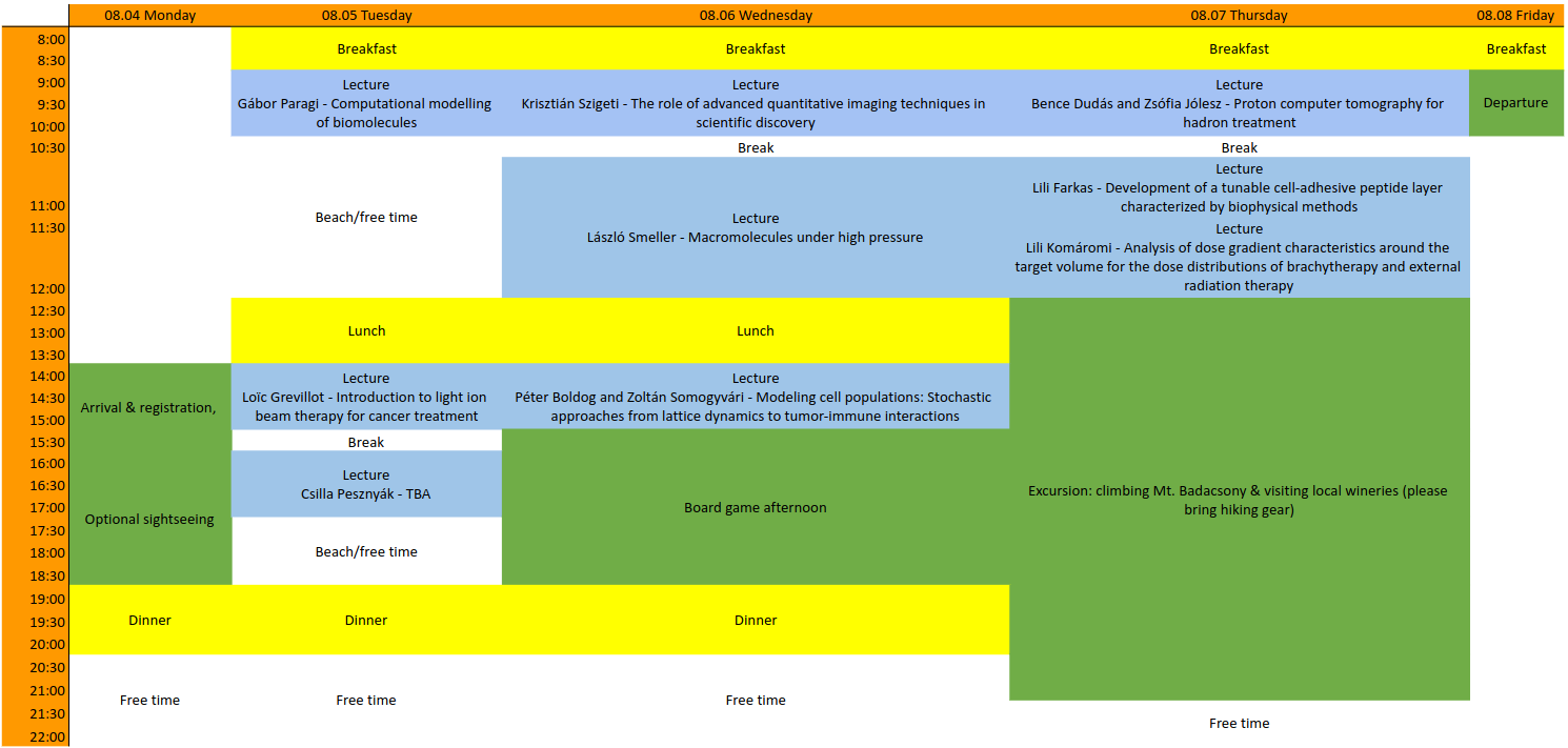
Lectures & Scientific Programme
Abstracts
Péter Boldog – Modeling cell populations: Stochastic approaches from lattice dynamics to tumor-immune interactions ▼
Understanding the dynamics of cell populations is central to many areas
of modern biology and medicine, including cancer progression, tissue
regeneration, and the design of personalized therapies. Mathematical
modeling offers powerful tools to uncover how collective behavior
emerges from local interactions such as cell movement, proliferation,
death, and biochemical communication.
A key spatial constraint in cell cultures is volume exclusion — the
simple but fundamental rule that no two cells can occupy the same volume
in space. This principle plays a critical role in how cell populations
grow and evolve, particularly in dense environments like tumors. To
accurately capture these dynamics, exact stochastic simulation
algorithms (SSAs), such as Gillespie’s method, provide a reliable
framework. Unlike deterministic models, they preserve discrete and
random events, making them especially useful when the number of cells is
low or stochasticity drives the system’s behavior.
We introduce two novel lattice-based SSA extensions: the Prompt Decision
Method (PDM) and the Reduced Rate Method (RRM). Both methods are exact,
incorporate volume exclusion, and support a broad set of biologically
relevant reaction types, including contact-inhibited and
contact-promoted reactions. While PDM samples and rejects non-executable
reactions, RRM computes adjusted reaction propensities based on local
cell density, making it more efficient in crowded scenarios.
To explore practical applications, we present a three-dimensional tumor
organoid–immune cell model, where tumor cells and T-cells are simulated
on separate interacting lattices. The model captures key experimental
phenomena such as T-cell infiltration and cytotoxicity, using real
parameter data from organoid–TEG T-cell co-culture experiments.
Finally, we discuss how programmed cell death and realistic cell cycle
modeling affects population dynamics. When the decomposition of cells or
the interdivision times has a deterministic length or follows empirical
distributions, a surprising synchronization of cells may
emerge—especially under spatial constraints. This cell cycle–induced
synchronicity could be exploited in cancer therapy, for instance, by
timing drug delivery to phases where tumor cells are most vulnerable.
The lecture will walk through the theoretical foundations, algorithmic
advances, and biological insights provided by these models—combining
mathematics, computation, and experimental relevance in the study of
complex cell systems.
Bence Dudás and Zsófia Jólesz – Proton computer tomography for hadron treatment ▼
Hadron therapy is a form of cancer therapy, where we aim to destroy those cancerous cells that are hard to reach with surgery. Since this kind of approach differs from the classical gamma radiation therapy, the tomography methods used for that are not sufficient enough for Hadron Therapy. Proton Computed Tomography (pCT) is developed to achieve more accurate results for this kind of treatment. During pCT a detector system measures incoming particles that are scattered on a phantom. Our aim is to reconstruct the trajectory of the primary particles, obtain their kinetic energies and scattering angles and from that create medical images.
Lili Farkas – Development of a tunable cell-adhesive peptide layer characterized by biophysical methods ▼
Controlling cell adhesion on biomaterial surfaces is key for improving implant integration and enabling spatial cell organization in tissue engineering.
In the research for my Master’s thesis, I developed a peptide coating with tunable cell-adhesive properties by optimizing a protocol to achieve stable, adjustable surface presentation. I evaluated the resulting layer using three label-free biophysical platforms — Epic BT biosensor, FluidFM, and HoloMonitor — to assess layer formation, mechanical attributes, and cell‑binding performance.
I will outline the protocol development process, describe the substrates used, and detail the measurement workflows.
Ultimately, this talk will guide you through my research journey and unveil key discoveries that emerged along the way.
Loïc Grevillot – Introduction to light ion beam therapy for cancer treatment ▼
Cancer is one the major cause of death globally. Light ion beam therapy (LIBT), also called particle therapy (PT), is an advanced form of radiation therapy (RT) for cancer treatments. While RT is used in more than 50% of all cancer treatments, LIBT represents less than 1% of all RT treatments. Proton therapy is the most spread out PT treatment technique, but heavier ions such as carbon ions offer additional biological advantages and sometimes the only treatment option for certain rare diseases and radiation resistant tumors. In this course, we will review the introduction of radiation in the field of medicine: from the discovery of X-ray by Wilhelm Röntgen in 1895, to the start of LIBT experiments at the Lawrence Berkley National Laboratory in the 1950’s. The focus will be put on current practical considerations of LIBT, providing understanding of the physics processes, radiation biology and clinical examples. Finally, future trends in the field of LIBT will be presented and discussed in light of the current state of the art and on-going developments.
Gábor Paragi – Computational modelling of biomolecules ▼
The computational modelling of molecules and their properties is a widely used and accepted tool in different research fields just like physics, chemistry or materials science. There is no different in biology-related sciences (biophysics, biochemistry, pharmacology, medicine), where someone try to understand phenomena at atomic or molecular level. During my talk, I would like to mentioned the most important ideas and methods which are often applied in computational modelling. Of course, the presentation cannot be complete in a single discussion, as it could be the topics of a whole semester, but trends and principles can perhaps be reviewed in a short talk.
László Smeller – Macromolecules under high pressure ▼
Pressure is a rarely used thermodynamical parameter. However, pressure experiments can provide such unique information about the macromolecules, which cannot be obtained otherwise. I will explain how to define and measure the volume of a solute macromolecule. I will present the theoretical description of the pressure-temperature phase diagram of proteins and other macromolecules, and introduce the experimental techniques used. Regarding experiments, besides the method of obtaining high pressure, I present the most frequently used spectroscopical techniques (IR, fluorescence) as well. I also talk about practical applications.
Krisztián Szigeti – The role of advanced quantitative imaging techniques in scientific discovery ▼
Medical imaging has become one of the most transformative tools in modern biomedical research and clinical diagnostics. This presentation explores how quantitative molecular and multimodal in vivo imaging techniques—such as PET, SPECT, MRI, CT, and functional ultrasound—are revolutionizing our understanding of complex diseases, including stroke, cancer, and neuroinflammation. We will explore the three foundational pillars that define the landscape of modern medical imaging. The first pillar is imaging modalities, which encompasses the physical principles behind techniques such as CT, MRI, and PET, as well as their hybrid combinations. These modalities differ significantly in how they visualize anatomical structures and physiological processes, offering complementary insights into the human body. The second pillar involves radioactive isotopes, which serve both diagnostic and therapeutic purposes. Radiotracers enable the visualization of molecular and cellular functions in vivo, providing a powerful tool for early disease detection and treatment monitoring. The third pillar is data evaluation, where advanced computational methods—such as radiomics, artificial intelligence, and big data analytics—are employed to extract meaningful patterns and biomarkers from the vast amount of imaging data generated. Together, these three elements form a synergistic framework that is transforming both research and clinical practice. Through real-world examples from stroke models and oncological applications, the talk will highlight how these technologies enable early detection, functional assessment, and personalized therapy planning. Special attention will be given to low-field portable MRI, targeted alpha therapy, and the integration of AI-driven image analysis. The goal is to inspire young physicists to see imaging not just as a diagnostic tool, but as a powerful research platform that bridges physics, biology, and clinical innovation.
Komáromi Lili – Analysis of dose gradient characteristics around the target volume for the dose distributions of brachytherapy and external radiation therapy ▼
Breast cancer is one of the most common types of cancer and requires constant development in treatment methods. In my presentation I am going to walk you through the basics of radiotherapy and show examples of its two types, teletherapy and brachytherapy. The goal of my Master’s research was to provide a simple, general method for decision making between these techniques.
The comparison between teletherapy and brachytherapy can be done by analyzing the dose profiles in all axes – lateral, medial, anterior, posterior, superior and inferior. After function fitting parametric evidence can be provided that there is significant difference between these methods, which gives opportunity to compare certain parameters of the plans. With the help of my method the dose of the organs at risk can be ultimately predicted easily which supports clinical decision making.
